Case Study

Atypical Spitz nevus versus Spitz melanoma
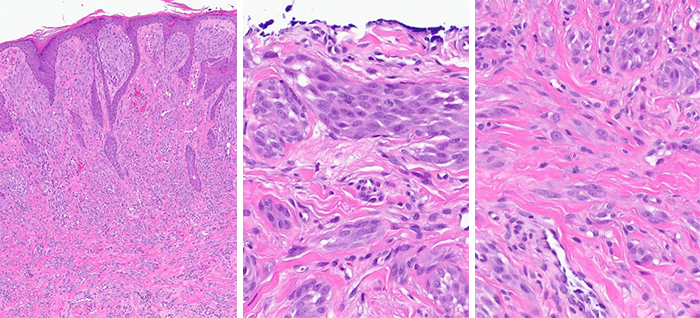
A 24-year-old male presents with a lesion on his left superior helix.
Case details

A 24-year-old male presents with a lesion on his left superior helix. A shave biopsy was performed.

- Compound melanocytic proliferation with enlarged spindled to epithelioid melanocytes mostly arranged in nests at the DEJ and in the dermis. Clefting around some of the nests is present. Focal confluent growth and minimal pagetoid spread are seen. Scattered deep mitotic figures are present.
- The lesion is transected at the deep and peripheral margins.
- Sox-10 staining highlights the melanocytes, revealing no confluence and only focal pagetoid scatter. p16 expression is retained. These findings are suggestive of a benign nevus.
- The melanocytes are however strongly positive (>75%) for PRAME suggesting malignancy.

The spitzoid morphology, retained p16 expression, and young age of the patient suggest that this lesion is a form of spitz nevus. However, the diffuse PRAME positivity and scattered deep mitotic figures are worrisome and could indicate a malignant process.
Given the high clinical stakes of missing or misdiagnosing a melanoma in a young patient, MyPath Melanoma GEP was ordered to help differentiate between an atypical Spitz nevus and a Spitz melanoma.
MyPath Melanoma gene expression profile resulted in a score of 2.7 which is suggestive of a malignant neoplasm.
A final diagnosis of a Spitz melanoma was given. With the aid of GEP testing, this patient received an accurate diagnosis of melanoma, ensuring the appropriate and timely treatment for this malignant lesion.